Intra-Oral Cameras – What Can You See? More Lesions – Part 5 – Bone Sequestra
Comparisons
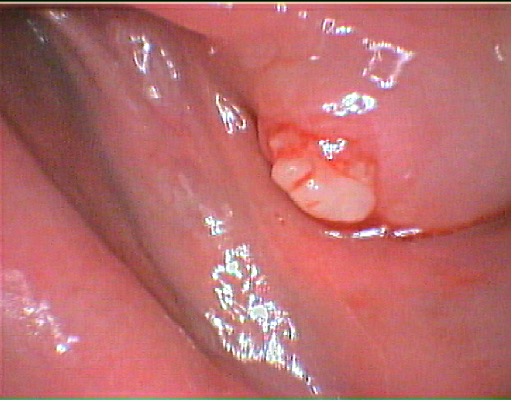
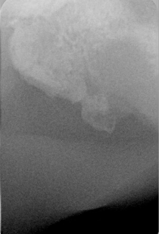
We often compare an x-ray image with the camera’s image. The camera shows how it lesion looks from the surface and the x-ray shows how it looks underneath.

An example of this is seen in bone sequestra.
This is a response by the body when a bit of bone has died. This can happen when a tooth is removed. In the course of removing the tooth, some bone may have been pushed aside and its blood supply has been cut off. This bone fragment eventually dies. The body responds by trying to “dissolve” it via its immune system as it is an irritant.
However, the body has another means of dealing with the fragment by pushing it out of itself through the gum. Hence, a bone sequestra forms.
There is a lump on the gum. There is a history of a recent extraction. So the camera is a highlighter of what’s going on in the mouth at on a grand scale, and because it was uncertain as to what the course was, an x-ray was taken to verify what was underneath.

The camera, with its own light, greatly enhances the image more than the naked eye, with magnification, as it shows up the texture, the colour and proportion in relation to other tissue.
Obviously it’s also an extremely important means of recording and detecting lesion growth or stability or healing status.
Treatment
Most of the time, the fragment is pushed out without the patient noticing it. Sometimes it’s painful if the dissolving bone fragment has forms a sharp edge. We then need to remove the fragment by directly removing it if it is exposed. Otherwise we need to open the gum to remove it and clean out any infections. A suture may be needed to close the gum for better healing outcomes.
Need an Appointment?
If you’d like to book an appointment with the dentist at Seymour Dental then call us in Dulwich Hill, Sydney on (02) 9564 2397 or
contact us
Next week
Easter 2024