Intra-Oral Cameras – What CanYou See? Bites and Grinds – Part 1
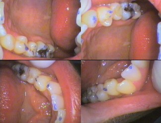
The intra-oral camera has been an incredible diagnostic tooth with its own light source. It has the ability to “see around corners” to area that are often missed by the naked eye, or even with magnification. The storing of images on the computer allows further technology to aid in diagnosis. The computer also enables the archiving of images and comparing past images for further diagnostic capabilities. Sharing of this with specialists and colleagues instantly for advice is also benefit for the dentist and therefore for the patient.
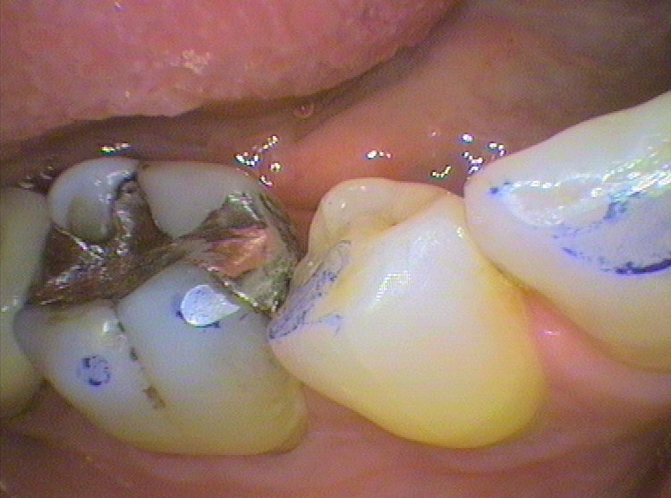
An assessment of the bite may show that it is uneven, causing the body attempt to even it by grinding teeth. This makes a flat shiny surface called a bruxofacet.


The comparison of before and after treatment adjustments can be viewed by patient. This allows the patient to be aware of where in the mouth was the problem.

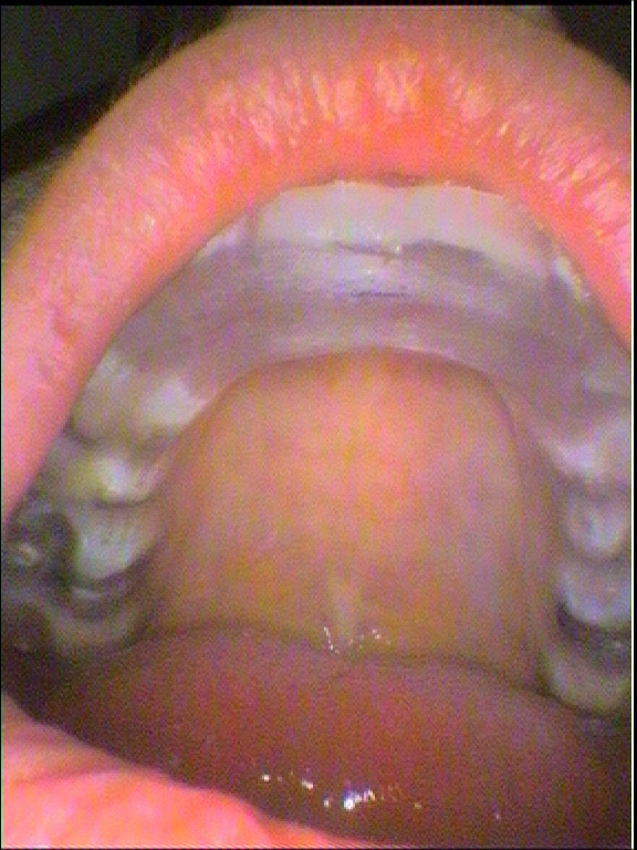
Sometimes making Occlusal splints to protects the teeth may be required. The camera can be used to check the pattern of the bite/wear and the fit. This is to that the fit is ccomfortable and to ensure the bite is still even. Always bring dentures, splints and other appliances for the dentist to check.

Next in series: Intra-Oral Cameras – What CanYou See? Bites and Grinds – Severe – Part 2
Need an Appointment?
If you’d like to book an appointment with the dentist at Seymour Dental then call us in Dulwich Hill, Sydney on (02) 9564 2397 or
contact us
Next week
Inala Art Exhibition 2023- After the Rain